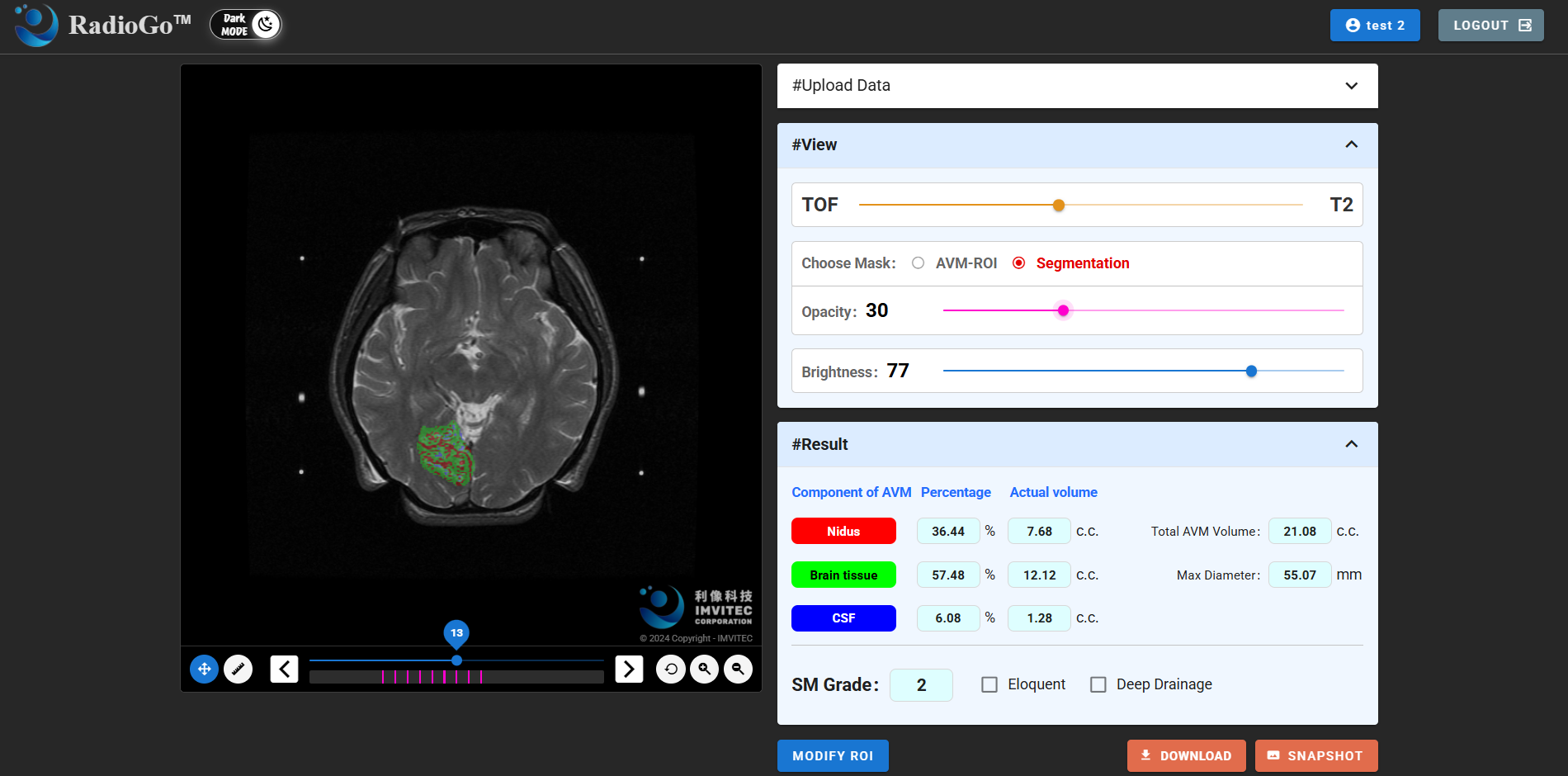
AI-AVM is designed to support the analysis of multimodal brain MRI images, particularly T2W and ToF sequences. The module assists with image registration, AVM lesion identification, segmentation of the nidus, brain tissue, and cerebrospinal fluid, as well as 3D volume calculation. These capabilities allow physicians to review lesion structures and surrounding anatomical relationships more efficiently before radiosurgery or Gamma Knife treatment.
Cerebral AVM is a complex neurovascular condition that often requires careful evaluation across multiple MRI sequences before treatment planning. In conventional workflows, physicians need to repeatedly compare different imaging sequences to identify the nidus, surrounding brain tissue, cerebrospinal fluid, and spatial anatomical relationships. This process can be time-consuming and highly dependent on clinical experience. AI-AVM is designed to transform these imaging findings into more consistent, quantitative, and visualized supportive information, helping clinical teams improve the efficiency of pre-treatment assessment.
Unlike standalone AI tools, AI-AVM is integrated into the broader RadioGo® AI-MRI platform. AI-MRI provides image ingestion, MRI sequence governance, lesion detection and localization, urgency and severity ranking, 2D and 3D visualization, report generation, and data governance. When a case requires disease-specific AVM analysis, physicians can switch to AI-AVM for advanced T2W/ToF registration, tissue segmentation, and 3D volume calculation.
For neurosurgeons, radiologists, and radiosurgery teams, the size, volume, and spatial relationship of the AVM nidus to surrounding brain tissue are important factors in treatment planning and risk assessment. By providing automated segmentation and 3D visualization, AI-AVM helps clinical teams better understand lesion structures. The system also supports standard export formats such as DICOM SEG, RTSTRUCT, and NIfTI, allowing analysis results to be further used in clinical review, research validation, and pre-treatment support workflows.

Current validation directions for AI-AVM include T2W/ToF image registration, segmentation of AVM nidus, brain tissue and CSF, clinical imaging validation, usability evaluation, and end-to-end system performance. According to the project document, AI-AVM tissue segmentation is evaluated using indicators such as Nidus Dice, Brain Dice, and CSF Dice, with DSC ≥ 80% set as an acceptance direction.
IMVITEC noted that World Brain Day reminds the public of the importance of brain health, prevention, and early awareness. At the same time, medical technology plays a critical role in supporting clinicians with more efficient and reliable imaging tools. RadioGo® AI-AVM is not intended to replace physician judgment. Instead, it provides AI-assisted image registration, tissue segmentation, and 3D volume calculation to generate faster, more consistent, and more quantitative imaging information for clinical review and treatment discussion.
Looking ahead, IMVITEC will continue to advance RadioGo® as a platform-based brain MRI imaging solution, combining broad imaging workflow capabilities with disease-specific AI modules. Through the development of intelligent medical imaging technologies, the company aims to support clinical teams in improving imaging review efficiency while echoing the spirit of World Brain Day: raising awareness of brain health and providing better tools to help protect every brain.